 Issue PDF
Editor’s note: In this Letter to the Editor, a reader details the challenges in maintaining adequate oxygenation and ventilation during upper GI endoscopy. This topic is related to the feature article on Non-Operating Room Anesthesia in this issue of the Newsletter.
Letter to the Editor
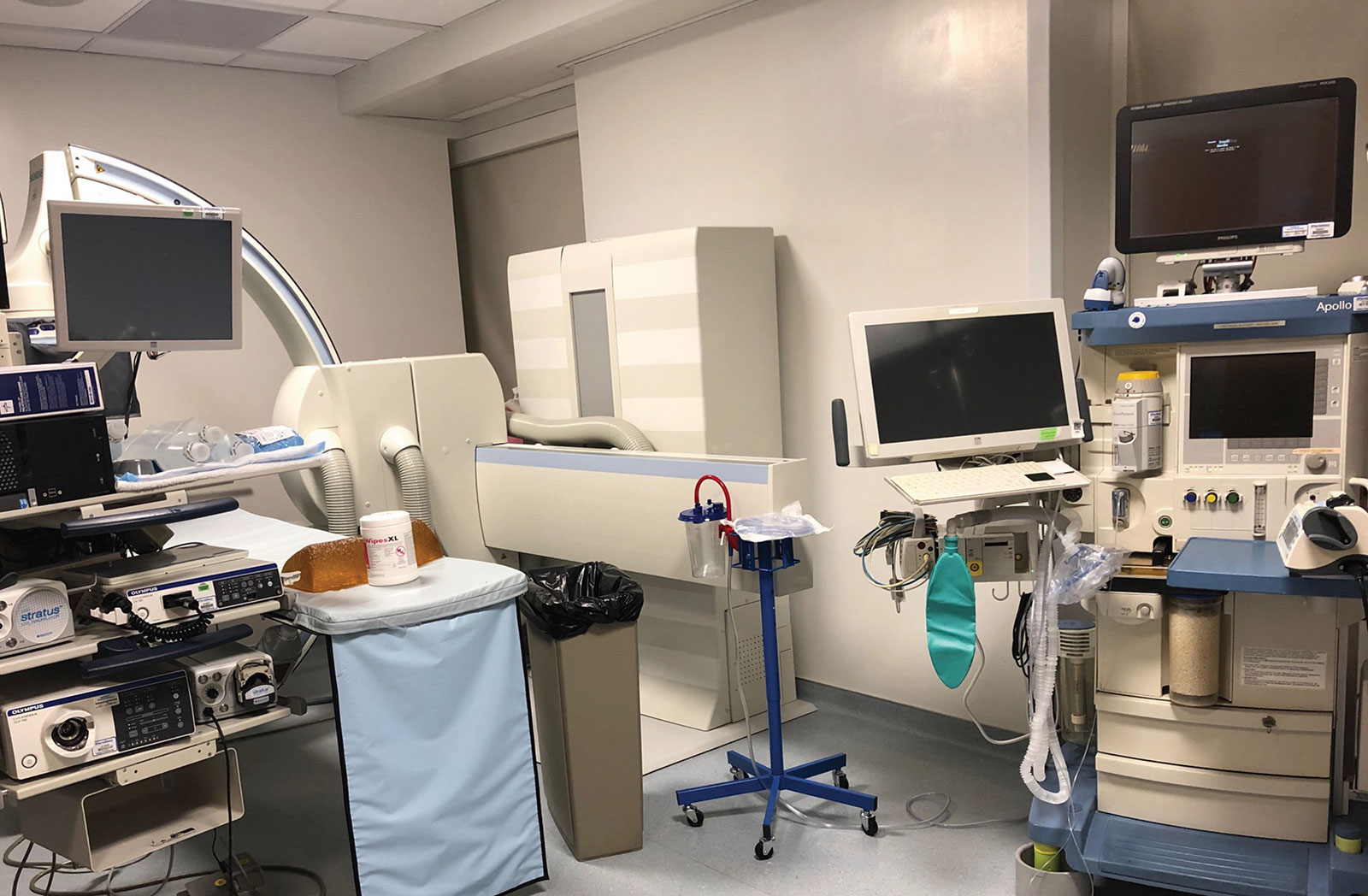
Endoscopy Suite
Upper endoscopies, even “simple” esophago-gastro-duodenoscopies (EGD’s), are challenging, potentially high-risk anesthetics for a number of reasons. They are by definition “shared airway” cases. They are also “reduced airway access” cases, since the patient is typically placed in the lateral, semi-prone, or prone position, greatly reducing the anesthesia professional’s access to the airway. Teeth can be dislodged by the bite block. Underlying pathologies (esophageal reflux, dysphagia, food impactions, GI bleeds, anemia, preparation for bariatric surgery) put these patients at risk for airway-related complications. Many are performed in procedure rooms with no anesthesia machine in case of an airway emergency and in locations remote from backup resources in the operating room. Rooms are darkened to allow the gastroenterologist a better view of the monitor screen. High case volume, quick case time, and room turnover pressures place time stress on the anesthesia professional, and can lead to the temptation to try to hurry the sedation, which can lead to “stacking” of doses of sedative agents, resulting in even deeper-than-desired levels of sedation. Computerized anesthesia records can create distractions for the anesthesia team, and can often force them to turn their back to the patient to face the computer screen.
Upper endoscopies often require very deep sedation bordering on general anesthesia to suppress the gag, cough, and laryngospasm reflexes, especially with initial insertion of the endoscope. Subsequently, the level of stimulation (and depth of sedation) can vary suddenly and significantly. Upper endoscopies are also, by definition, foreign body obstruction cases, since a large foreign body, the endoscope, is placed into the aero-digestive tract, often producing partial airway obstruction. Sedation reduces muscle tone of the upper airway, which may result in airway collapse that anesthesia professionals must commonly manage.1
The diameter of commonly used adult esophago-gastroscopes is 8.8-11 mm.2 If we apply the formula for the area of a circle, A= π r2, it becomes evident that the cross-sectional area of a 9 mm endoscope often exceeds the cross-sectional area of the airway documented on CT studies,3,4 thereby presenting a risk for partial or even total obstruction of the airway in a significant percentage of patients. The Guardus® Overtube (US Endoscopy, Mentor, OH), a clear plastic tubular device placed over the endoscope to create aerodigestive separation for removal of impacted food, has an even larger outer diameter of 19.5 mm.5
Another major challenge in delivering sedation for upper endoscopy has been the limitation of our supplemental oxygen delivery systems. Traditional oxygen facemasks for upper endoscopy are typically not used as they impair access to the mouth by the endoscopist. Often, the mode of oxygen delivery is one of our least effective: nasal cannula (or,insufflation via an oral catheter).
Standard nasal cannulae are recommended to be used at maximal O2 flows of 5–6 L/min.6 Even short durations of higher rates are not well tolerated because of discomfort and drying of the nares that may result in epistaxis. At O2 flows of 6–7 L/min, nasal cannulae provide a maximum FiO2 of approximately 0.44–0.62.6 Other common clinical conditions, such as nasal congestion, nasal polyps, or septal deviation can further reduce the oxygen delivery from a nasal cannulae. By contrast, O2 face masks with non-rebreathing reservoirs, at O2 flows of 9–15 L/min, comfortably provide much higher FiO2’s of approximately 0.90–0.95.
Given the issues of airway encroachment and the potential for limited oxygen delivery, airway management during upper endoscopy under sedation is, by its very nature, “high risk.” Therefore, anesthesia professionals should approach these cases similar to the way we approach patients requiring general anesthesia in the operating room—by remembering the time-tested principles of “safe apneic time” and “maximal preoxygenation.”
“Safe apneic time” is defined as the delay from the onset of apnea until the SpO2 drops to below 90%, into the steep portion of the hemoglobin-O2 desaturation curve and into critically low levels. The safe apneic time in healthy adults is approximately less than one minute.7 However, patients with decreased capacity for oxygen loading (e.g., anemia, pulmonary disease, obesity, decreased cardiac output, or decreased functional residual capacity), or with increased oxygen demand (fever, hypermetabolic state) desaturate much more quickly.8-10
It has been established for decades that the simple technique of “maximal preoxygenation” can double or even triple safe apneic time.7-9 In a classic 1999 editorial in Anesthesiology, Dr. Jonathan Benumof wrote: “The purposes of maximally preoxygenating before the induction of general anesthesia are to provide the maximum time that a patient can tolerate apnea, and for the anesthesia professional to solve a cannot-ventilate/cannot intubate situation. Moreover, because a cannot ventilate/cannot intubate situation is largely unpredictable, the desirability to maximally preoxygenate is theoretically present for all patients.”10 Dr. Benumof vigorously espoused maximal preoxygenation whenever possible. Preoxygenation has become standard practice for many practitioners prior to all general anesthetic inductions (i.e., iatrogenically-induced apnea).8,9
There are several accepted methods of effective maximal preoxygenation.10,11 Many techniques used by anesthesia professionals require O2 flow (>10L/min) through a well-fitting oxygen mask. The most effective and efficient may be the “8 DB/60 sec” (8 deep breaths over 60 seconds) method described by Baraka.10,11 Dr. Benumof’s logic, which has served our patients so well for decades in the potentially high-risk situation of induced apnea in the operating room, should be extended to patients undergoing upper endoscopy under sedation, particularly if this can be done simply and cost-effectively.
In recent years, there have been several oxygen masks designed specifically for upper endoscopy procedures.1 These oxygen masks deliver reliably high oxygen concentrations while providing capnography monitoring capabilities and easy endoscopic access.
Capnography and vigilance allow rapid diagnosis of severe hypoventilation, even in a dark room with the patient facing away from us. There are now available endoscopy oxygen facemasks and other devices that make the goal of providing near-maximal preoxygenation prior to the start of deep sedation attainable. These devices may prolong safe apneic time to allow intervention prior to the onset of severe hypoxia.1
Improving the safety of the patients we serve requires continual re-assessment of our practices, and a willingness to improve where possible. Since 1955, we have had a simple method, “maximal preoxygenation,” available to prolong safe apneic time.12 In 2019, there is equipment available enabling us to approximate “maximal pre-oxygenation” prior to induction of hypopnea and insertion of an obstructive endoscope into the upper airway. In his “2019 President’s Report,” Dr. Mark Warner, reiterated the APSF’s vision that “no patient shall be harmed by anesthesia,” and implores us all to continue to work on “this noble quest.”13 Therefore, our goal should be zero tolerance for hypoxia during upper endoscopies. There is room and opportunity for improvement.
René Miguel Gonzalez, MD, is a staff anesthesiologist at Hackensack Meridian Southern Ocean Medical Center.
The author has no conflicts of interest pertaining to this article.
References
- Goudra B, Singh PM. Airway management during upper GI endoscopic procedures: state of the art review. Dig Dis Sci. 2017;62:45–43.
- Flexible endoscope general diameter guide. Available at: www.healthmark.info/Flexible_Scopes_General_Diameter_Guide.pdf. Accessed February 25, 2019.
- Avrahami E, Solomonovich, Englender M: Axial CT measurements of the cross-sectional area of the oropharynx in adults with obstructive sleep apnea syndrome. Am J Neuroradiol. 1996;17:1107–1111.
- Li H, Lo Y, Wang C, et al. Dynamic drug-induced sleep computed tomography in adults with obstructive sleep apnea. Scientific Reports 2017. Available at: www.nature.com/scientificreports. Accessed February 25, 2019.
- Guardus overtube. Available at: www.usendoscopy.com/products/guardus-overtube. Accessed February 25, 2019.
- Wettstein R, Shelledy D, Peters J. Delivered oxygen concentrations using low-flow and high-flow nasal cannulas. Respiratory Care. 2005;50:604–609.
- Drummond GB, Park GR. Arterial oxygen saturation before intubation of the trachea—an assessment of techniques. Br J Anaesth. 1984;56:987–9.
- Bouroche G, Bourgain JL. Preoxygenation and general anesthesia: a review. Minerva Anestesiol. 2015;81:910–920.
- Sirian R, Wills J. Physiology of apnoea and the benefits of preoxygenation. Continuing Education in Anaesthesia Critical Care & Pain. 2009;9:105–108.
- Benumof JL. Editorial views. Preoxygenation: best method for both efficacy and efficiency? Anesthesiology. 1999; 91:603–605.
- Baraka AS, Taha S, Aouad M, et al. Preoxygenation: Comparison of maximal breathing and tidal volume breathing techniques. Anesthesiology. 1999;91:612–616.
- Hamilton W, Eastwood D. A study of denitrogenation with some inhalational anesthetic systems. Anesthesiology. 1955;16:861–867.
- Warner, MA. 2019 President’s report: taking action. APSF Newsletter. 2019;33:69–71. https://dev2.apsf.org/article/2019-presidents-report-taking-action-apsfs-renewed-commitment-to-implementation-of-changes-that-can-improve-perioperative-patient-safety/. Accessed April 8, 2019.